Eye drops
Pressure-lowering drops are common first-line treatment. Technique, side effects and adherence should be reviewed.
Shifa International Hospital, Islamabad
Optic nerve care
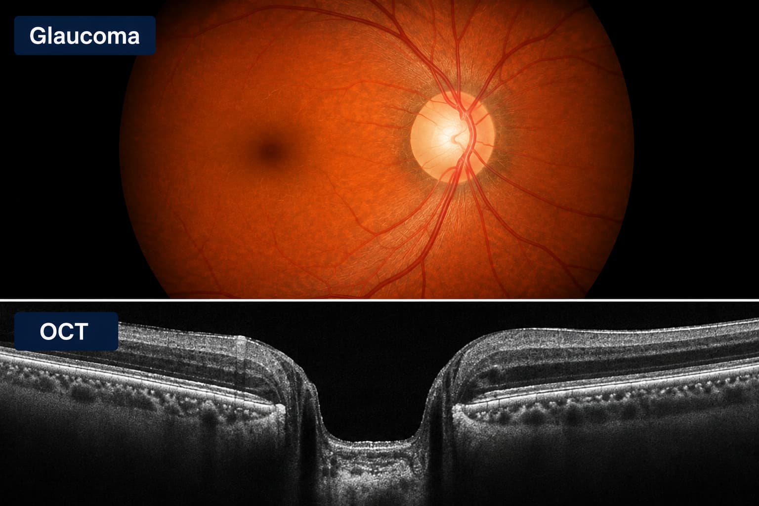
Glaucoma can damage the optic nerve gradually and without early symptoms. Examination combines pressure measurement with optic-nerve and visual-field assessment.
Sudden severe eye pain, headache, nausea, coloured halos, redness or rapid visual loss require urgent in-person assessment.
Understanding the concern
Glaucoma describes a group of conditions that damage the optic nerve. Eye pressure is an important risk factor, but glaucoma can occur at different pressures and a single pressure reading cannot confirm or exclude it.
Open-angle glaucoma often progresses silently. Regular examination is especially important for people with a family history, increasing age, diabetes, high myopia, steroid exposure or previous eye injury.
What patients notice
These features can overlap with other eye conditions. Examination is needed to establish the cause.
Specialist assessment
A glaucoma review combines several measurements over time rather than relying on one test.
Pressure is measured and interpreted alongside corneal thickness and other ocular findings.
Examination and imaging look for structural change in the optic nerve and retinal nerve fibre layer.
A field test maps side vision and can help identify or monitor functional damage.
Gonioscopy helps distinguish open from narrow drainage angles and informs treatment planning.
Treatment planning
Treatment is tailored to glaucoma type, stage, pressure, rate of change and the patient's ability to use medication consistently.
Pressure-lowering drops are common first-line treatment. Technique, side effects and adherence should be reviewed.
Laser may be used to improve drainage or treat narrow angles in selected types of glaucoma.
When drops and laser are insufficient or disease is advanced, a glaucoma surgical pathway may be recommended or coordinated with a glaucoma subspecialist.
Optic nerve assessment
At a glance
Prof. Amer Awan's wider ophthalmic practice includes glaucoma assessment, while complex or progressive glaucoma may require coordinated subspecialty management according to the examination findings.
Questions patients ask
Usually not. Most open-angle glaucoma causes no early pain or warning symptoms, which is why examination and testing are important.
No. Optic-nerve susceptibility varies, and glaucoma can occur at statistically normal pressure. Diagnosis uses the full clinical picture.
Established optic-nerve damage is generally permanent. Treatment aims to reduce the risk of further loss, making early detection valuable.
Continue reading
General information sources: Prof. Amer Awan - current public profile; National Eye Institute - Facts about glaucoma.