Observation
Some very early or partial changes can be monitored with repeat OCT when spontaneous improvement remains possible.
Shifa International Hospital, Islamabad
Macular surgery
A macular hole affects the central retina and may cause distortion, a blurred central patch or difficulty with detail.
Understanding the concern
The macula is responsible for detailed central vision. A full-thickness macular hole is a small opening in this tissue, usually related to traction as the vitreous changes with age, although trauma and other factors can also contribute.
OCT imaging confirms the diagnosis and shows the size and shape of the hole. Observation may be appropriate in selected early situations, while established holes are commonly discussed in relation to vitrectomy surgery.
What patients notice
These features can overlap with other eye conditions. Examination is needed to establish the cause.
Specialist assessment
Assessment establishes whether there is a full-thickness hole, associated vitreous traction and any other macular condition affecting the likely benefit of treatment.
OCT provides a cross-sectional view of the macula and is central to confirming and measuring a macular hole.
The rest of the retina is examined for tears, detachment, epiretinal membrane and other disease.
Cataract status and the vitreomacular relationship in the other eye may influence counselling and planning.
Treatment planning
The recommendation depends on hole stage, size, symptom duration, visual impact and the condition of the eye.
Some very early or partial changes can be monitored with repeat OCT when spontaneous improvement remains possible.
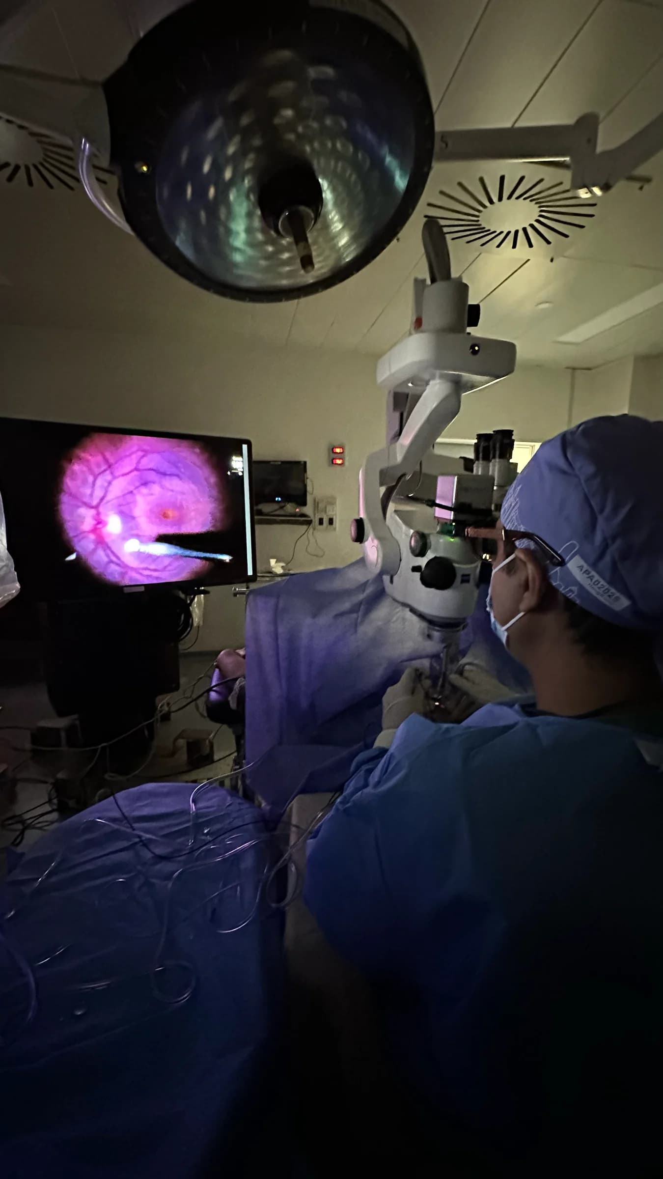
Surgery removes vitreous traction and typically involves careful peeling of a fine internal limiting membrane around the macula.
A gas bubble may support closure while the eye heals. The type and duration of positioning are determined by the operative plan.
Macular surgery
At a glance
Prof. Amer Awan's vitreoretinal work includes macular-hole assessment and small-gauge surgery. OCT findings, symptoms and the health of the eye guide whether observation or vitrectomy is appropriate.
Related surgical video
From Prof. Amer Awan's official YouTube channel. This professional video contains surgical footage.
Questions patients ask
No. Both affect the macula, but a macular hole is an opening related to tissue traction, while age-related macular degeneration is a different disease process.
Glasses can optimise focus but do not close a full-thickness hole. OCT examination is needed to determine the stage and options.
Positioning recommendations vary with hole size, surgical technique and surgeon preference. Follow the instructions given for your operation.
Continue reading
General information sources: Prof. Amer Awan - current public profile.