Observation
Mild, stable symptoms can be followed with repeat vision checks and OCT imaging.
Shifa International Hospital, Islamabad
Macular conditions
An epiretinal membrane is a fine layer of tissue on the macula that can wrinkle the retina and distort central vision.
Understanding the concern
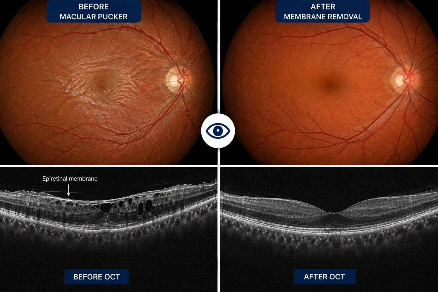
A macular pucker, also called an epiretinal membrane, forms on the surface of the macula. Mild membranes can be found incidentally and may not need treatment. Others cause increasing distortion, blur or difficulty using the two eyes together.
OCT imaging shows the membrane and its effect on retinal structure. The decision to observe or operate is based on symptoms and function rather than the scan appearance alone.
What patients notice
These features can overlap with other eye conditions. Examination is needed to establish the cause.
Specialist assessment
The examination distinguishes an epiretinal membrane from macular hole, swelling, AMD and other causes of central distortion.
OCT maps the membrane, traction and retinal thickness and provides a baseline for monitoring.
Visual acuity and patient-reported distortion help establish how much the membrane affects daily tasks.
The peripheral retina, lens and other macular structures are reviewed before any treatment decision.
Treatment planning
Not every membrane needs treatment. Surgery is considered when symptoms are significant enough to justify the risks and recovery.
Mild, stable symptoms can be followed with repeat vision checks and OCT imaging.
The vitreous is removed and the membrane is carefully peeled from the macular surface. Visual improvement can be gradual and may be incomplete.
Vitrectomy can influence cataract progression. Lens status and any cataract symptoms are considered as part of the surgical discussion.
Macular assessment
At a glance
Prof. Amer Awan's vitreoretinal work includes epiretinal membrane assessment and macular surgery. The decision to monitor or operate depends on distortion, visual function, OCT findings and the likely benefit for the individual eye.
Related surgical video
From Prof. Amer Awan's official YouTube channel. This professional video contains surgical footage.
Questions patients ask
It usually develops gradually and is not typically an emergency. Sudden loss of vision, flashes, many new floaters or a curtain still require urgent assessment for other retinal problems.
No. Glasses may optimise focus, but the membrane itself is only removed surgically. Mild cases often do not need removal.
No complete outcome can be promised. Improvement is often gradual and depends on how long the retina has been distorted and its underlying health.
Continue reading
General information sources: Prof. Amer Awan - current public profile.